Imaging criteria for UCAS Japan
The UCAS Japan study includes unruptured aneurysms larger than 3 mm in diameter. We consider the accurate measurement of the aneurysm's size to be particularly important for drawing meaningful conclusions.
We strongly advise all participating researchers to use a unified standard to measure aneurysms. A brief summary of the method is given here.
Diagnosis
- The diagnosis of an unruptured intracranial aneurysm should be made by board certified neurosurgeons or radiologists (Japan Neurosurgical Society, Japan Radiological Society).
- Images used for the diagnosis should be obtained through one of the following methods:
- Routine angiography
- Digital subtraction angiography (DSA)
- Three-dimensional computed tomographic angiography (3D-CTA)
- Magnetic resonance angiography (MRA) obtained with an MRI machine with a magnetic field of more than 0.5 Tesla
Size measurement
The size of the aneurysm should be defined according to the largest diameter of the aneurysm measured by using one of the following methods:
- Digital imaging such as 3D-CTA, MRA, or MRI
- Angiography (DSA or routine) obtained with size-references such as grids or a 1-yen coin (the 1-yen coin method)
- In patients with a thrombosed aneurysm, the outer diameter of the aneurysm is regarded as the aneurysm's size and should be measured by using 3D-CTA.
Accuracy of diagnosis and size measurement
The accuracy of the diagnosis and the size measurements are checked periodically at randomly selected institutions by qualified examiners sent by the central administrative office.
Measurement of the size of the aneurysms: 1-yen coin method
Since digital subtraction angiography or other conventional angiography inevitably produce image distortion and magnification, we need to provide uniform method to calculate magnification ratio of acquired angiography at each institution. There are several method to calculate magnification ratio and lesion size, such as catheter tip calibration method, rectangular grid phantom placement and the 1-dime coin method utilized in ISUIA. Catheter calibration method can induce significant measurement error between investigators. Other materials, rectangular grid phantom or one dime coin are not usually available in Japanese institutions.
 A 1-yen coin, universally available in Japan, is exactly 20 mm in diameter
and is made of aluminum, which can be penetrated by diagnostic X-rays. The
coin's image can be seen on X-ray pictures.
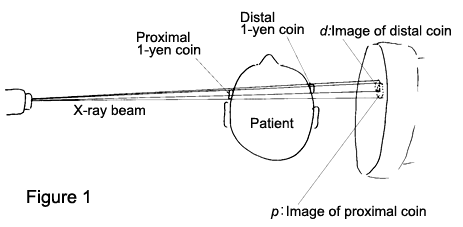
By placing 1-yen coins as easily available references on both sides of the head along the X-ray trajectory, we can calculate the magnification at the center of the head.
This method is recommended for unruptured intracranial aneurysms measured with angiography machines that do not have size references.
Steps
Before obtaining images of the target aneurysm, place 1-yen coins along the X-ray trajectory. The first one is placed on the patient's head nearest the imaging equipment (proximal). The second coin is placed on the other side of the patient's head (distal). At the time of angiography, scout pictures should be obtained (Figure 1).   |
|
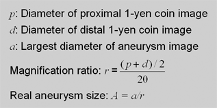
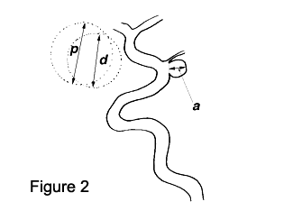
On the scout picture, measure the largest diameter of the 1-yen coin images at the front and rear of the skull (p & d). The average of these sizes divided by 20 mm is the magnification ratio (r) of the image (Figure 2).  |
|
| Measure the largest diameter of the aneurysm on the image (a). Divide this number by the magnification ratio (r) to find the real size of the aneurysm (A). |
Accuracy of this 1-yen coin method was cross verified with Grid phantom method and was shown to be a reliable method before included in the protocol.


